Re-engineering of acetyl coenzyme A metabolism prevents senescence in budding yeast

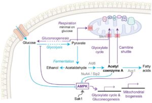
A diagram showing aspects of metabolism in a yeast cell, emphasizing glycolytic flux leading to acetyl coenzyme A and pathways promoted by AMPK.
Metabolic pathways acting for and against senescence

For many, the idea of living forever is unappealing. Even if good health is thrown into the bargain, interest in immortality remains ambivalent. But what if we could stay healthy right up until a timely death? Recent work from the Houseley lab suggests that, at least in budding yeast, healthspan can be extended without also extending lifespan. In a 2023 paper, they showed that a galactose diet suppresses senescence (i.e., loss of fitness) in aging yeast without extending their lifespan. In a recent preprint, they claim to achieve the same effect in yeast on a normal (glucose-rich) diet by activating AMPK while simultaneously preventing AMPK-mediated repression of fatty acid synthesis.

Here’s an intriguing sentence from the discussion (that, however, might benefit from being split into two sentences): “It is not too surprising that issues arise when AMPK is ectopically activated on high glucose given that AMPK is a central energy regulator that evolved to manage metabolism under low nutrient conditions, and it is perhaps more surprising that a single point mutation largely corrects these issues, providing optimism that the benefits of AMPK activation could also be maximised in higher eukaryotes.”


Re-engineering of acetyl coenzyme A metabolism prevents senescence in budding yeast
bioRxiv, March 2025
From the lab of Jon Houseley.

Snippet by Katrina Woolcock.

Image credit: Figure 5 from Hadj-Moussa et al. linked above (CC-BY).

Microbial ecosystems and ecological driving forces in the deepest ocean sediments

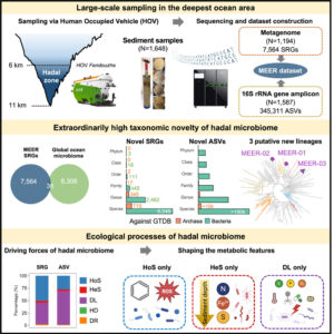
A complex diagram of the methods and results of the paper, depicting the vehicle, sampling apparatus, taxonomic distribution of organisms, and the kinds of processes that have driven their evolutionSome scientific articles are must-reads because of what the scientists accomplished or developed. Some scientific articles are must-reads because of what the scientists discovered. And then some scientific articles are must-reads for both reasons. Here’s an example from a few weeks ago. A group of scientific teams in China worked for years to develop a vehicle that could carry researchers to Hell and back, so they could bring back dirt and animals from Hell. Then they went to Hell, brought back soil, and extensively analyzed the microbial communities in Hell. They discovered that these communities are dramatically distinct from communities in the nearby ocean, and thereby illuminated surprising evolutionary trajectories of the microbial organisms that inhabit Hell.

I’m overdramatizing. The scientists didn’t visit Hell. They visited Hades, which is mythologically different. Okay fine, their inspiring and successful mission went to the hadal zone of the ocean (named, aptly, after the dark underworld of Hades), specifically to the mysterious famous chasms of the Mariana Trench that are nearly 11,000 meters (about 6.8 miles) deep. Somehow, amazingly, microbes can live and reproduce and evolve at that depth. Maybe they’re even happy.

Here is the abstract from their Cell paper. I think it’s a bit too understated, but it is inspiring nonetheless, and we can all use some inspiration right now.

Systematic exploration of the hadal zone, Earth’s deepest oceanic realm, has historically faced technical limitations. Here, we collected 1,648 sediment samples at 6–11 km in the Mariana Trench, Yap Trench, and Philippine Basin for the Mariana Trench Environment and Ecology Research (MEER) project. Metagenomic and 16S rRNA gene amplicon sequencing generated the 92-Tbp MEER dataset, comprising 7,564 species (89.4% unreported), indicating high taxonomic novelty. Unlike in reported environments, neutral drift played a minimal role, while homogeneous selection (HoS, 50.5%) and dispersal limitation (DL, 43.8%) emerged as dominant ecological drivers. HoS favored streamlined genomes with key functions for hadal adaptation, e.g., aromatic compound utilization (oligotrophic adaptation) and antioxidation (high-pressure adaptation). Conversely, DL promoted versatile metabolism with larger genomes. These findings indicated that environmental factors drive the high taxonomic novelty in the hadal zone, advancing our understanding of the ecological mechanisms governing microbial ecosystems in such an extreme oceanic environment.


Microbial ecosystems and ecological driving forces in the deepest ocean sediments

In Cell, 6 March 2025 (this issue of Cell includes three papers and a Commentary piece on the hadal ocean research)
From the groups of Xiang Xiao, Weishu Zhao, Wei-Jia Zhang, Mo Han, Xun Xu, and Shanshan Liu

Snippet by Stephen Matheson

Image credit: graphical abstract from Xiao et al. linked above (CC-BY)

De novo designed proteins neutralize lethal snake venom toxins

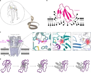
The figure shows the structure of a peptide toxin, emanating from a drawing of a cobra. Other panels illustrate how the toxin interacts with the membrane and with nicotinic ACh receptors.
A cobra and its toxin: Figure 1 from Vázquez Torres et al.

Can you name a neglected tropical disease (NTD)? I would guess that most biologists can, or can at least make some accurate guesses. But by definition, these are maladies that don’t get adequate attention and resources, and so it is likely that many scientists are unaware of the current roster of NTDs. A new and exciting paper at Nature taught me that snakebite (envenoming, to be precise) is in fact a devastating NTD, with a heartbreaking death toll due in large part to the lack of effective and affordable* treatments. More importantly, the paper reports on how the authors discovered some simple and effective therapeutics with significant potential to make a difference. Their abstract is, to me, a tour de force of scientific writing that is straightforward and inspiring. (My favorite sentence suggests that we can “democratize therapeutic discovery.”) Here it is with references removed for readability:

Snakebite envenoming remains a devastating and neglected tropical disease, claiming over 100,000 lives annually and causing severe complications and long-lasting disabilities for many more. Three-finger toxins (3FTx) are highly toxic components of elapid snake venoms that can cause diverse pathologies, including severe tissue damage and inhibition of nicotinic acetylcholine receptors, resulting in life-threatening neurotoxicity. At present, the only available treatments for snakebites consist of polyclonal antibodies derived from the plasma of immunized animals, which have high cost and limited efficacy against 3FTxs. Here we used deep learning methods to de novo design proteins to bind short-chain and long-chain α-neurotoxins and cytotoxins from the 3FTx family. With limited experimental screening, we obtained protein designs with remarkable thermal stability, high binding affinity and near-atomic-level agreement with the computational models. The designed proteins effectively neutralized all three 3FTx subfamilies in vitro and protected mice from a lethal neurotoxin challenge. Such potent, stable and readily manufacturable toxin-neutralizing proteins could provide the basis for safer, cost-effective and widely accessible next-generation antivenom therapeutics. Beyond snakebite, our results highlight how computational design could help democratize therapeutic discovery, particularly in resource-limited settings, by substantially reducing costs and resource requirements for the development of therapies for neglected tropical diseases.

*In a world in which a corrupt oligarch can buy the executive branch of the US government using less than 1% of his wealth, let’s not pretend that the main problem is “affordability.” The authors describe several other reasons why current approaches are badly inadequate.


De novo designed proteins neutralize lethal snake venom toxins
In Nature, 15 January 2025
From the groups of Timothy P. Jenkins and David Baker.

Snippet by Stephen Matheson

Image credit: Figure 1 from Vázquez Torres et al. linked above (CC-BY-NC-ND).

A plant virus manipulates both its host plant and the insect that facilitates its transmission

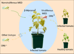
A tomato plant is in the middle. From the left, a fly approaches, labelled "nonviruliferous." On the plant, another fly is colored red and is now viruliferous (what we're calling infected). This fly departs (indicated by arrows) to infect another plant, pictured on the right. The attractant and the olfactory receptor are indicated by text labels.
Figure 6 from Liang et al.

Viruses and other parasites are famously adept at trickery and manipulation. Think of your favorite creepy example of that, then consider a new paper about a virus so devious that it might become your new favorite (if you’re into that sort of thing). The article in Science Advances reveals the strategic brilliance of the tomato yellow leaf curl virus (TYLCV), which infects tomato plants with the help of its vector, a whitefly. Step 1 of the evil plan: the virus induces the plant to make a protein that attracts the whitefly. Nice move, but now there’s a new problem, which is that flies are attracted to already-infected plants. Our scheming virus wants the flies to carry it to uninfected plants. So, step 2: the virus inhibits the fly’s olfactory receptor that makes it home in on that attractive signal. At first this might seem like the virus is undoing its work, but think about it: now only uninfected flies will be attracted to the infected plants. That means more infected flies, which now no longer prefer the infected plants! [virus voice] MWAAAHAHAHAHA.

Here the authors nicely summarize the evil plan, using vivid language that highlights the strategic brilliance of this devastating pathogen:

These results reveal a unique two-tiered pull-push strategy in which TYLCV maximizes transmission by modulating terpene release from host plants as well as the olfactory system of its vector.


A plant virus manipulates both its host plant and the insect that facilitates its transmission
In Science Advances, 28 February 2025
From the groups of Ted Turlings, Wen Xie, and Youjun Zhang.

Snippet by Stephen Matheson

Image credit: Figure 6 from Liang et al. linked above (CC-BY-NC-ND).

Chromosome-scale genome assembly reveals how repeat elements shape non-coding RNA landscapes active during newt limb regeneration

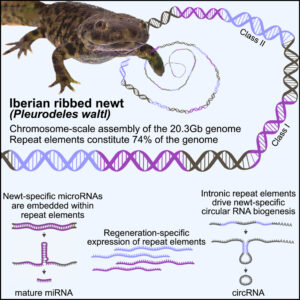
A diagram showing the newt (a brownish salamander), an artistic DNA strand, and labels describing the genome's features.
Graphical abstract from Brown et al.

Have you heard about the Iberian ribbed newt? This creature could be a vision from the Locked Tomb series by Tamsyn Muir. It’s goth, it’s metal, it’s… well look, if you are foolish enough to piss this animal off, it will stab you with its own ribs, which it brandishes through its skin and which gather poison on the way out. You will learn your lesson, and the newt will go on with her/his day, because it is also famous for its regenerative capacities.

The authors of a new paper in Cell Genomics undertook the epic quest to assemble the complete gigantic genome of this adorable nightmare. (Apparently the newt is more welcoming to genomic parasites than it is to human handlers.) The scientists seem more interested in regeneration than in gothic horrors, and they show that some of the newt’s vast hordes of genomic parasites (transposable elements) are likely involved in regeneration. The hordes are indeed vast: 3/4 of this animal’s giant genome is made of transposable elements. Here is their final paragraph, which nicely expands our vision beyond the newt and its gothness:

From a broader perspective, the data uncover how repeat elements shape a gigantic genome, serving as common denominators between genome expansion, facilitation of circRNA formation, and evolution of miRNAs from stem-loop structures. Furthermore, the chromosome-scale assembly of P. waltl provides a useful resource that facilitates our understanding of vertebrate chromosome evolution, not the least, variations among syntenic blocks and gene content. Our results thus allow for the exploration of species-specific innovations and evolutionarily shared molecular responses to major injuries, knowledge necessary to identify mechanisms that promote or counteract successful regeneration.


Chromosome-scale genome assembly reveals how repeat elements shape non-coding RNA landscapes active during newt limb regeneration
In Cell Genomics, 12 February 2025
From the labs of Nicholas D. Leigh, Maximina H. Yun, and András Simon.

Snippet by Stephen Matheson

Image credit: graphical abstract from Brown et al. linked above (CC-BY)

Hierarchical representations of relative numerical magnitudes in the human frontoparietal cortex

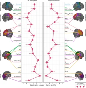
Two mirror-image graphs showing relative performance of classifying relative numerosity in various brain regions, which are depicted as pseudocolored images.
Relative numerosity coding in various brain regions.

Our brains detect and analyze myriad features of the world, and we all know that it does much of this work automatically. One thing it can do, seemingly without effort, is detect how many things are in a set or collection. (This isn’t counting, which is different.) The ability is called “numerical competence” and is widespread in animals that include insects but apparently not the unelected trolls who are firing people at NSF and NIH. Seriously, numerical competence is important for survival and reproduction, and the feature being detected is called numerosity.

A new paper in Nature Communications looks in detail at the brain regions and systems that detect numerosity, specifically asking how our brains detect absolute numerosity and then move to detecting/computing relative numerosity. The authors show that absolute numerosity is detected in the first stages of processing in the visual system, but that even at that early step, the brain is estimating relative numerosity. As processing moves into other “later” brain regions, relative numerosity is the focus. In their Discussion, the authors summarize this finding and suggest that it might apply to other wondrous capacities of our underused brains:

By illustrating that the representation of relative numerosity emerges through this hierarchical process, our study goes beyond these previous studies and introduces an additional dimension of abstraction: the transition from absolute to relative numerical magnitude. We speculate that this abstraction process might be a fundamental principle in magnitude coding, potentially offering more efficiency and robustness, and might even be applicable across other domains.


Hierarchical representations of relative numerical magnitudes in the human frontoparietal cortex
In Nature Communications, 6 January 2025
From the labs of Yuko Yotsumoto and Masamichi J. Hayashi.

Snippet by Stephen Matheson

Image credit: Figure 3 from Kido et al. linked above (CC-BY-NC-ND)

The microglial response to inhibition of Colony-stimulating-factor-1 receptor by PLX3397 differs by sex in adult mice

I sometimes make fun of microglia, because they were (unfairly) given a name that means “tiny glue.” They deserve better: these are immune cells that live in the brain and attend to numerous important matters (none of which involve glue). A new paper in Cell Reports shows that microglia in mice are strikingly different between males and females, and for me this adds to their mystique as quiet heroes of the brain and to the pathos of their silly name.

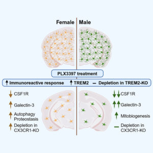
The illustration shows two halves of a brain in cross section, male and female, before and after treatment with the drug. The male brains have fewer remaining cells after treatment.
Graphical abstract from the paper

So, what is strikingly different? PLX3397 is an inhibitor of the CSF1 receptor, but what matters to us mere mortals is that its action causes loss of microglia from a treated brain. The drug is used widely to erase microglia, facilitating subsequent experimental manipulations. The authors show that its effects differ based on sex and that the difference is likely connected to very different actions of the surviving microglia: in males, the survivors make more mitochondria; in females, the survivors invest in protein retention and recycling (proteostasis and autophagy). As the authors conclude in their abstract: “These findings suggest sex-dependent microglial survival mechanisms, which might contribute to the well-documented sex differences in various neurological disorders.” That is an interesting and important result. But one more thing. The title was, I think, written for specialists who know a lot about CSF1 and who know what PLX3397 does. Those excluded from this company discover the key difference at the end of the title, which (please humor me) could have been “Sex differences in microglia are revealed by differential response to inhibition of Colony-stimulating factor.” Just a thought.


The microglial response to inhibition of Colony-stimulating-factor-1 receptor by PLX3397 differs by sex in adult mice
In Cell Reports, 25 January 2025
From the lab of Ania K. Majewska.

Snippet by Stephen Matheson

Image credit: graphical abstract from Le et al. linked above (CC-BY-NC-ND)

Anti-icing properties of polar bear fur

Polar bears are classic examples of “charismatic megafauna,” finishing a solid 8th in a 2018 survey of the most charismatic species on earth. (The Top 20 ranking includes zero birds, and 4 of the top 7 are murderous cats, which reveals more about humans than it does about other animals.) I certainly agree that they are charismatic, having watched them perform at a zoo many years ago, though I admit my fanhood was diminished by watching them hunt baby belugas in the not-very-deep blue sea.*

One thing that stands out is that fur. It’s not actually white—it’s hollow and thereby provides insulation. Maybe you knew that, but did you know that polar bear fur doesn’t accumulate ice? It should (water and cold and all) but it doesn’t, and a recent paper in Science Advances explains why: the fur contains a lot of greasy secretions. NPR already did a great job on the story, in which the lead author connects her team’s findings to people and the past: “We didn’t discover it,” she says. “It’s been known to Arctic people for centuries.” Their abstract notes that the work “builds on Inuit knowledge of natural anti-icing materials.”

In that vein, here is some nice writing in the Introduction, that caught my eye for its opening of a window to the past:

…polar bears are known to slide on snow and ice (Fig. 1A). An early account of this behavior is given by the Inuk hunter and artist Jakob Danielsen (1888 to 1938). He writes: The (polar bear) mother plays a lot with her young. We see in the mountains that they have made slides on the snow slopes down which they go on their tail all in the same track (25) (p. 317).

See bottom for a picture of a bear doing that! It’s Figure 1 of the paper.

*Listen to “Baby Beluga” by Raffi if my phrasing seems weird to you.


Anti-icing properties of polar bear fur
in Science Advances, 29 January 2025
From the lab/group of Bodil Holst.

Snippet by Stephen Matheson.

A polar bear is sliding down a snowbank, head first on her/his back. Other panels show a pseudocolored infrared image of a polar bear, and two other photos of bears shaking off ice and rolling on the snow.
Properties of polar bear fur in the wild. From Figure 1 of Carolan et al., cited above.

Tissue clocks derived from histological signatures of biological aging enable tissue-specific aging predictions from blood

A spiral made of DNA emerges, with 12 hours indicated by images of different tissues and organs.
Tissue clocks. Image by Gloria Fuentes.

Is age really just a number? Hundreds of “aging clocks” developed over the last 10–15 years suggest that it is. These clocks aim to measure biological age rather than chronological age—initially using DNA methylation, more recent examples use, e.g., blood proteins. A recent preprint goes a step further by developing “tissue clocks” that predict the biological age of different human tissues from images or even blood samples. The authors reason that tissue structure, with its direct links to physiological fitness, is a better indicator of aging than molecular and cellular changes. For example, dwindling blood vessel numbers and accumulating fibrosis (scarring) are tell-tale signs of tissue aging—wrinkles are not just skin-deep… Perhaps we will soon be able to test which of our organs are aging faster than others, taking personalized medicine to a new level.


Tissue clocks derived from histological signatures of biological aging enable tissue-specific aging predictions from blood
bioRxiv, November 2024
From the lab of André F. Rendeiro.

Snippet by Katrina Woolcock.

Single-cell data reveal heterogeneity of investment in ribosomes across a bacterial population

Consider whether you would read an article that reached a conclusion like this: “Our results thus reveal a range of strategies for investing resources in the most expensive machines at the heart of this process.” I probably wouldn’t, since words like “invest” and “expensive” are—shall we say—unattractive to me. But what if the sentence is actually this: “Our results thus reveal a range of strategies for investing resources in the molecular machines at the heart of cellular self-replication.” That’s a sentence from the abstract of this interesting paper in Nature Communications, “Single-cell data reveal heterogeneity of investment in ribosomes across a bacterial population.” The authors looked at ribosome numbers and synthesis in individual bacterial cells and found surprising diversity in ribosomal mass and in the timing of responses of individual cells to challenges. Populations are known to respond to metabolic challenges with corresponding shifts in allocation of resources to ribosomal synthesis, but the new paper shows that individual cells vary dramatically in how they do this.

The authors end their paper with this clear and compelling overview, brimming with great metaphors:

…we thus quantified, using a combination of reporter genes and statistical inference algorithms, dynamic investment in ribosomes on the single-cell level. The results reveal a surprising variability in the allocation of resources to ribosomes, the most costly molecular machine in bacterial cells, during both balanced and unbalanced growth. This raises fundamental questions on the role of the variability of ribosome concentrations in shaping the growth of a bacterial population and its adaptation to changing environments. Given the importance of growth and adaptation in biomedical and biotechnological applications, we expect our findings to have practical implications as well.


Single-cell data reveal heterogeneity of investment in ribosomes across a bacterial population
Nature Communications 2 Jan 2025
From the labs of Johannes Geiselmann and Hidde de Jong.

Snippet by Stephen Matheson